Captions and Attributions
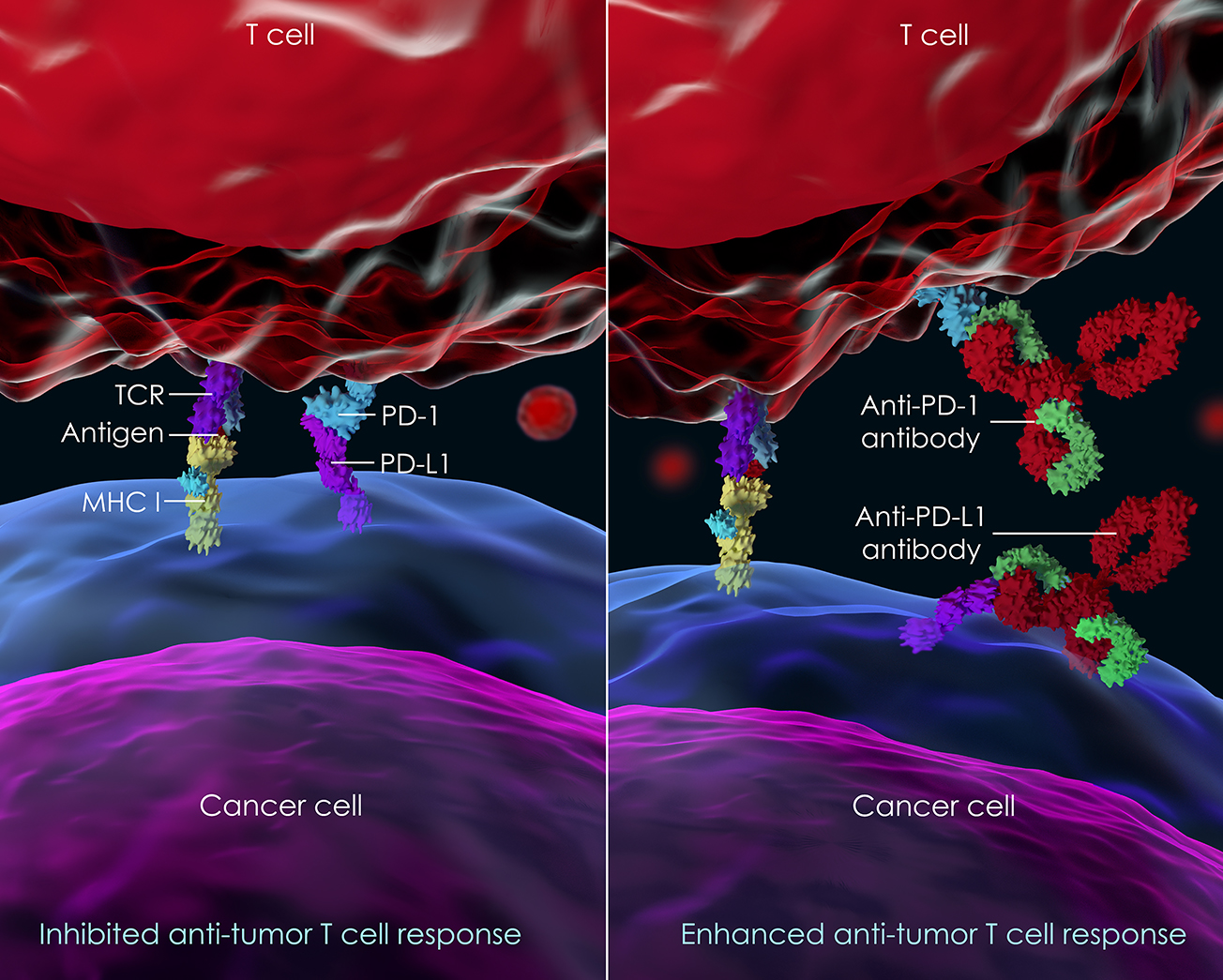
Figure 1: Blocking cell surface PD-1 with an immune checkpoint inhibitor (ICI) to enhance T cell antitumor activity. The left side of the image shows a cytotoxic T-Cell (red; a type of immune cell) adjacent to a cancer cell targeted for destruction (blue). Engagement of the cell surface T-cell receptor (TCR) with a cancer antigen complexed with MHC-1 is one of 2 initial steps that must occur for the T-cell to lyse the cancer cell. But here, a cell surface protein on the cancer cell, PD-L1, engages a T-cell receptor known as PD-1. This engagement sends a negative signal into the T-cell blocking the destruction of the cancer cell. But on the right, introduction of antibodies to block either PD-1 or PD-L1 (both examples of ICIs) blocks the interaction of PD-1 and PD-L1, removing the inhibitory signal into the T-cell and allowing the T-cell to destroy the cancer cell. Photo credit: iStock©
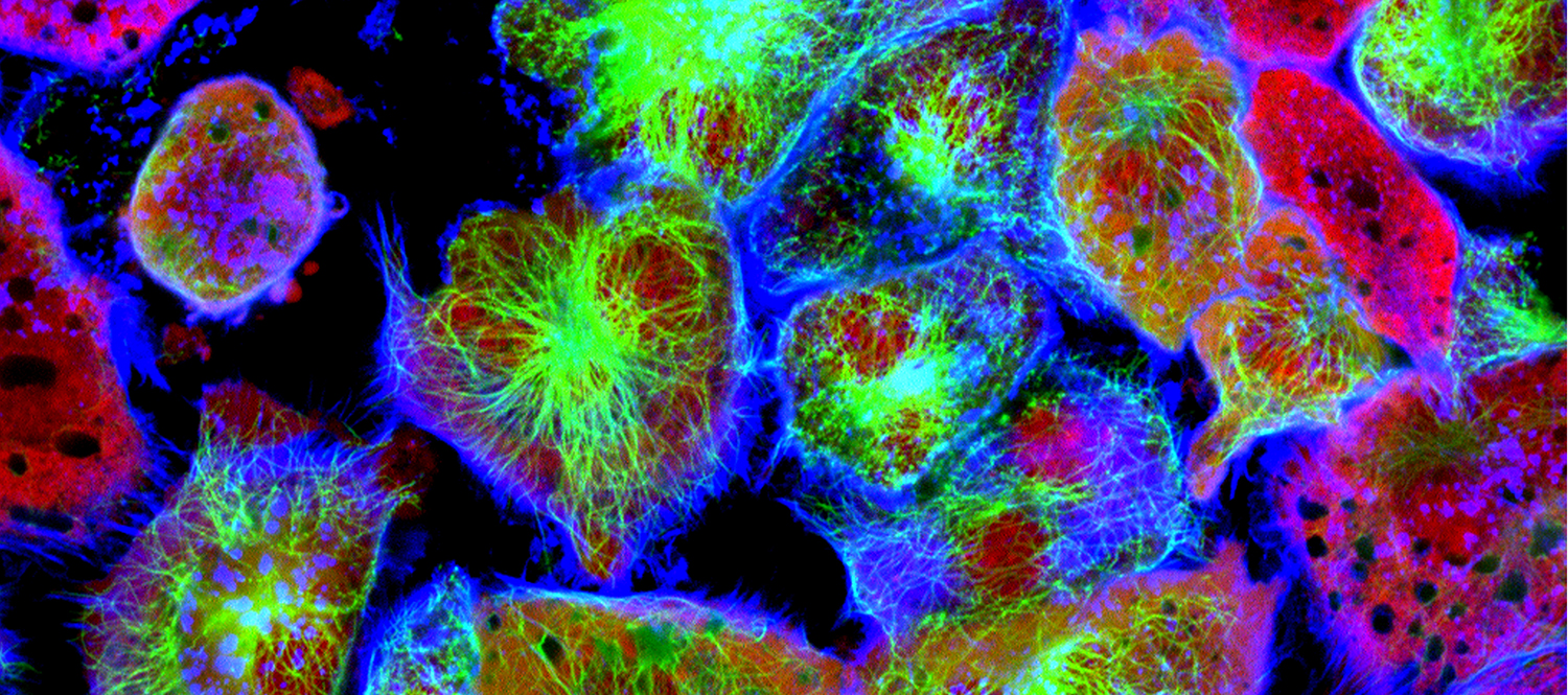
Figure 2: Assorted types of tumor cells labeled with different fluorescent probes. Note the striking diversity of cell types present in a tumor. Photo credit: iStock©
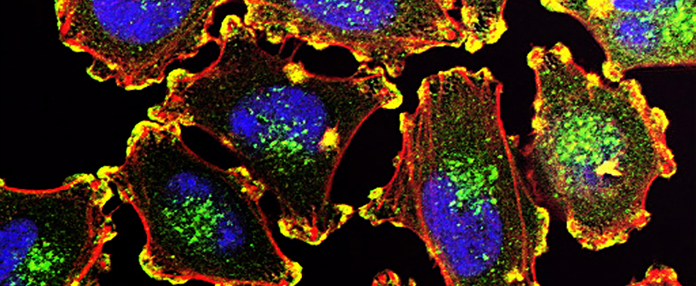
Figure 3: Metastatic Melanoma Cells. The ability of cancer cells to move and spread depends on the TME as well as actin-rich core structures such as the podosomes (yellow) shown here in melanoma cells. Cell nuclei (blue), actin (red), and an actin regulator (green) are also shown. Photo Credit: National Cancer Institute on Unsplash.
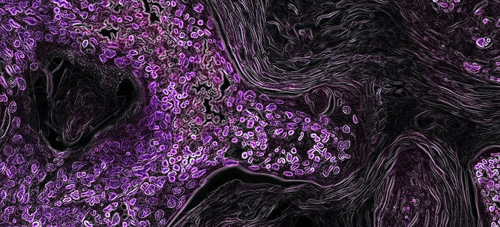
Figure 4: KRAS-Driven Lung Cancer. One of the great success stories of modern cancer therapy is the use of ICIs to treat some types of lung cancer patients. Photo credit: By Eric Snyder, 2015, National Cancer Institute on Unsplash.
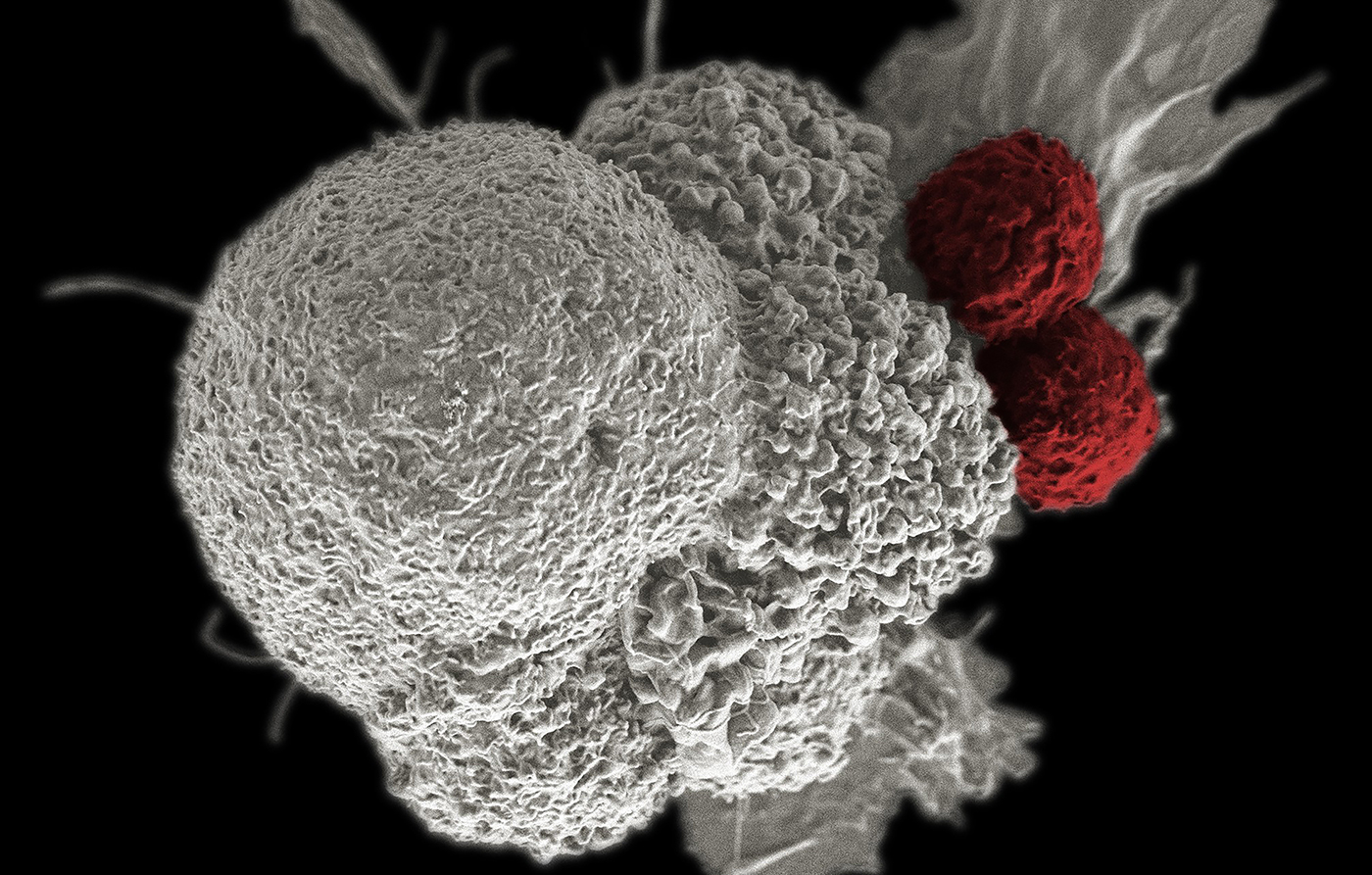
Figure: Cancer Immunotherapy - A pseudo-colored scanning electron micrograph of an oral squamous cancer cell (white) being attacked by two cytotoxic T cells (red). Photo credit: By Rita Elena Serda, 2015, National Cancer Institute \ Duncan Comprehensive Cancer Center at Baylor College of Medicine on Unsplash.